Benign Paroxysmal Positional Vertigo (BPPV) is a common vestibular disorder that causes brief episodes of vertigo triggered by changes in head position.
Among the various types of BPPV, Horizontal Canal BPPV (HC-BPPV) is particularly challenging to diagnose and treat. The supine roll test, also called Pagnini-Lempert test or Pagnini-McClure Roll Test, is a crucial diagnostic tool for identifying HC-BPPV.
This blog covers:
- What HC-BPPV is
- Contradictions for the supine roll test
- How to perform the roll test
- Interpreting results
- Understanding the underlying mechanisms
- HC-BPPV treatment
- Video demonstration of the supine head roll test
Understanding Horizontal Canal BPPV
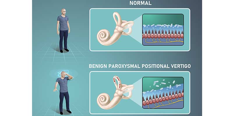
Benign paroxysmal positional vertigo from the lateral semicircular or simply horizonal canal BPPV, occurs when otoconia, or calcium carbonate crystals, dislodge from the utricle and enter the horizontal semicircular canal.
This displacement causes abnormal canal stimulation during head movements, leading to vertigo and nystagmus. Head trauma and ear infections are common causes of HC-BPPV, though the majority of cases are idiopathic, meaning they occur without a known cause.
Benign Paroxysmal Positional Vertigo affects the lateral semicircular canal in approximately 5-15% of cases. The roll test helps identify the affected ear and the type of nystagmus, essential for effective treatment.
Contraindications for the Roll Test
Before performing the supine roll test, consider these contraindications to ensure patient safety:
- Severe Neck or Spine Injuries: Avoid the test if the patient has severe neck or spine injuries.
- Acute Neurological Symptoms: Stop the test if symptoms like confusion, numbness, blurred vision, or limb weakness occur.
- Recent Head Trauma: Be cautious with patients who have recently experienced head trauma.
- Severe Cardiovascular Conditions: Patients with uncontrolled hypertension or recent heart issues should not undergo the test.
- Severe Anxiety or panic disorders: The test may trigger anxiety or panic attacks in susceptible individuals.
Performing the Supine Roll Test
The roll test is performed with the patient in a supine position. Here are the steps to conduct the test:
1. Preparation: Explain the procedure to the patient and ensure they are comfortable. Then, position the patient supine position with their head elevated 30 degrees to align the horizontal canal with the plane of gravity.
2. Initial Position: Start with the patient's head in a neutral position. Observe for any spontaneous nystagmus, which may indicate a central vestibular disorder.
3. Head Movement to the Affected Side: Turn the patient's head 90 degrees to one side (right or left) and observe the eye movement. This head movement stimulates the horizontal canal on the turned side.
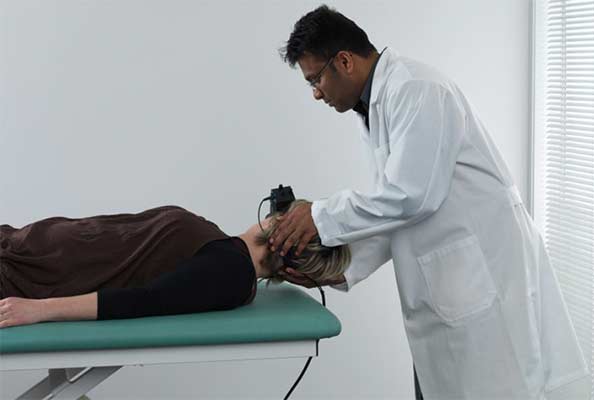
Head roll to the right.
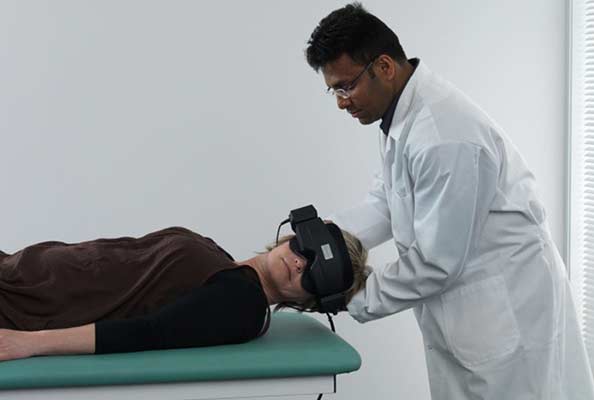
Head roll to the left.
4. Observing Nystagmus: Look for horizontal nystagmus. If geotropic nystagmus (towards the ground) is observed, it indicates canalithiasis, where the otoconia are free-floating in the canal. If apogeotropic nystagmus (away from the ground) is observed, it suggests cupulolithiasis, where the otoconia are attached to the cupula.
5. Returning to Neutral: Return the patient's head to the neutral position and allow nystagmus to subside.
6. Head Movement to the Opposite Side: Turn the patient's head 90 degrees to the opposite side and observe for nystagmus again. Compare the intensity and direction of nystagmus on both sides.
Interpreting the Roll Test Results
The roll test helps determine the affected ear and the type of HC-BPPV:
- Geotropic Nystagmus: If the nystagmus is stronger when the head is turned towards one side, that side is the affected ear. Geotropic nystagmus indicates canalithiasis.
- Apogeotropic Nystagmus: If the nystagmus is stronger when the head is turned away from one side, the opposite side is the affected ear. Apogeotropic nystagmus indicates cupulolithiasis.
Treatment Based on Roll Test Results
Once the affected ear and type of HC-BPPV are identified, appropriate treatment maneuvers can be performed:
- For Geotropic Nystagmus (Canalithiasis), the BBQ Roll maneuver or the Gufoni maneuver can be used to move the otoconia out of the horizontal canal.
- For Apogeotropic Nystagmus (Cupulolithiasis), The Gufoni maneuver or the Modified Semont maneuver can be effective in dislodging the otoconia from the cupula.
For instructions on how to treat HC-BPPV, including the BBQ roll maneuver and Gufoni manuever, please refer here: Horizontal Canal BPPV: Diagnosis and Treatment
VNG and Diagnosing Horizontal Canal BPPV
Videonystagmography (VNG) goggles are an essential diagnostic tool for HC-BPPV. By capturing eye movements during positional changes, these goggles enable clinicians to analyze the direction and characteristics of nystagmus. This detailed observation aids in identifying the affected canal and selecting the most suitable treatment.
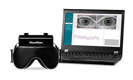
Interacoustics Visual Eyes 505 VNG goggles
Learn More
Practical Tips for Physical Therapists
- Patient Comfort: Ensure the patient is comfortable and informed throughout the procedure. Sudden head movements can be distressing, so proceed gently.
- Observation Skills: Accurate observation of eye movements is crucial. To enhance the visibility of nystagmus, use Frenzel goggles or videonystagmography.
- Documentation: Record the intensity, direction, and duration of nystagmus for both head positions. This information is vital for diagnosis and treatment planning.
- Follow-Up: After performing the treatment maneuvers, reassess the patient to ensure the symptoms have resolved. Educate the patient on home exercises and precautions to prevent recurrence.
The supine roll test is an essential diagnostic tool for identifying Horizontal Canal BPPV. Physical therapists can effectively manage HC-BPPV and improve patient outcomes by understanding the procedure, interpreting the results, and applying appropriate treatment maneuvers.
Do you want to learn more about diagnosing and treating balance disorders with advanced equipment? Our local experts are here to assist you.
Contact Local Expert
Other Good Reads: What’s the Difference Between BPPV and Cervical Vertigo?
Watch Now: How to Diagnose Lateral Canal BPPV
Follow us on Social!


If you haven't already, make sure to subscribe to our newsletter to keep up-to-date with our latest resources and product information.