
Practical oVEMP Testing

Many clinics that do diagnostic vestibular testing will benefit from adding Vestibular Evoked Myogenic Potential (VEMP) recordings to the battery of tests they perform on patients with dizzy/balance issues. Adding VEMP testing, along with the Video Head Impulse Test (vHIT), can provide a more comprehensive look into the function of the individual components of the vestibular system and, therefore, greatly improve the sensitivity of the test battery for detecting vestibular disorders.
The VEMP tests are relatively quick and easy to perform, even on patients with sensorineural hearing loss. These tests can be conduced on some of the most popular Auditory Evoked Potential (AEP) instruments and may not require a significant additional investment. This article will focus on the Ocular Vestibular Myogenic Potential (oVEMP) test.
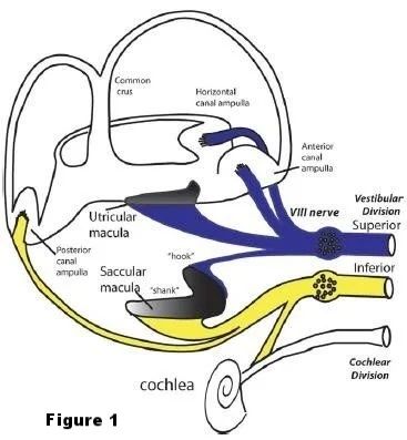
The oVEMP response is generated by the utricle and is mediated by the superior vestibular nerve (See Figure 1). It is recorded on the oblique ocular muscle. The oVEMP is the result of the synchronous evoked extraocular muscle activity associated with the VOR and does not represent movements of the eyes (i.e. is not EOG). Unlike the cervical cVEMP, which originates in the saccule and is mediated by the inferior vestibular nerve, the oVEMP is an excitatory, not inhibitory, muscle response.
The same pathologies that can affect the cVEMP (peripheral vestibular impairments) can also affect the oVEMP. It depends on which anatomical structures are affected. With pathology involving the associated anatomical structures, contralateral responses are typically reduced or absent. The following disorders many be indicated:
- Vestibular Neuritis
- Acoustic Neuroma
- Labyrinthitis
- Brainstem Stroke
- Meniere’s Disease
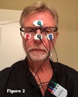
We have been successful using the electrode montage listed below:
Inverting (-) electrodes under each eye
Non-inverting (+) electrode on the nose
Ground electrode on the forehead (See figure 2).
We also use an AEP test protocol that is similar to the one we use for the cVEMP, except that the gain is much higher. (See below.) - 500 Hz Tone Burst
- 2-0-2 cycle rise-fall time
- Linear gating
- Rep Rate = 5 – 8 /sec
- Stimulus level = 95+
- Gain = 100,000 or 75,000
- Window = approx. 45 mSec
- Gaze – up (in order to move the oblique m. closer to the electrode)
- Artifact reject = 40uV
- Filter = 10 to 400 Hz
- Samples = 300 to 400
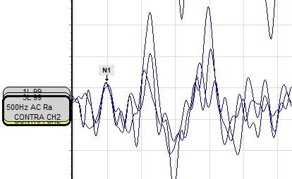
Note that the patient is told to look upward and to continue gazing upward as long as he or she hears the stimulus. With the electrode montage indicated above, the first significant positive going part of the response (N1) occurs at approximately 10ms, and the first significant negative going part of the response occurs at approximately 14ms.
The N1 response should be repeatable over two or more repeated recordings as shown below.
Many clinics will reverse the waveforms in order to conform to criteria used in the majority of published studies. Figure 3 shows the waveforms reversed and marked accordingly.
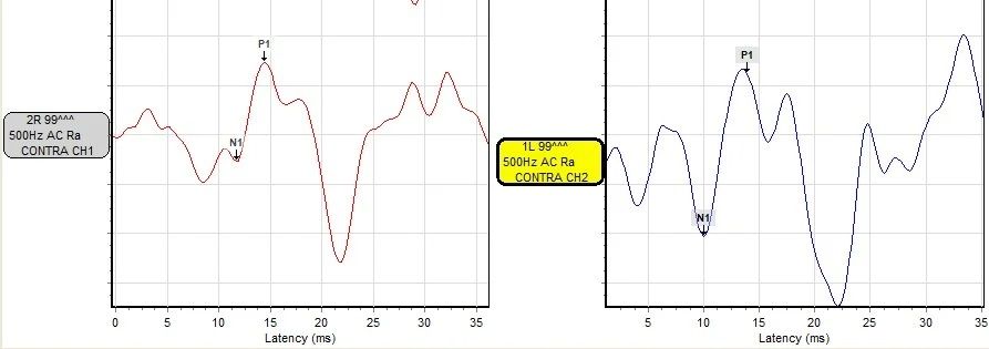
Figure 3
With peripheral vestibular disorders, the N1 response is often reduced or missing. The oVEMP is considered abnormal when the asymmetry ratio of the left and right N1-P1 amplitude exceeds .35. The amplitude asymmetry ratio is simply the difference of the N1-P1 absolute amplitudes (in microvolts) in the right and left contra recordings over the sum of the same N1-P1 absolute amplitudes (in microvolts).
In the example shown above, the right contra N1-P1 amplitude is .8 microvolts and the left N1-P1 amplitude is 1.32 microvolts. After calculating the difference over the sum, the left-right amplitude asymmetry ratio is .245 which is well within normal.
- N1 = approx. 10 mSec
- P1 = approx. 14 mSec
- Response is recorded from the contralateral eye.
- N1-P1 amplitude asymmetry ratio is abnormal when it exceeds .35
VEMP testing (both cVEMP and oVEMP) can be a valuable asset for comprehensive vestibular assessment. With the addition of VEMP recordings as well as vHIT, all major components of the vestibular system are tested: all 6 semi-circular canals as well as the saccule, the inferior vestibular nerve, the utricle and the superior vestibular nerve in each ear.